I have just made my annual pilgrimage to Oxford, seat of brainy scientists and medieval architecture. It was Meeting Minds 2026, where I keenly attend all the medical lectures while sheepishly admitting “I did history actually” to anyone who tries to engage me in any semi-scientific conversation.
I booked a room in Magdalen College which is both beautiful and impressive, but the dodgy electrics and bathroom facilities have made me consider an IBIS for next year. I am just feeling a bit too old and a bit too “21st century” to find elusive, malfunctioning light switches, and a creepy climb up a long flights of wooden stairs for a midnight pee break, in any way romantic. And yes, I think if I’m writing about Oxford I’m allowed to use an Oxford comma.
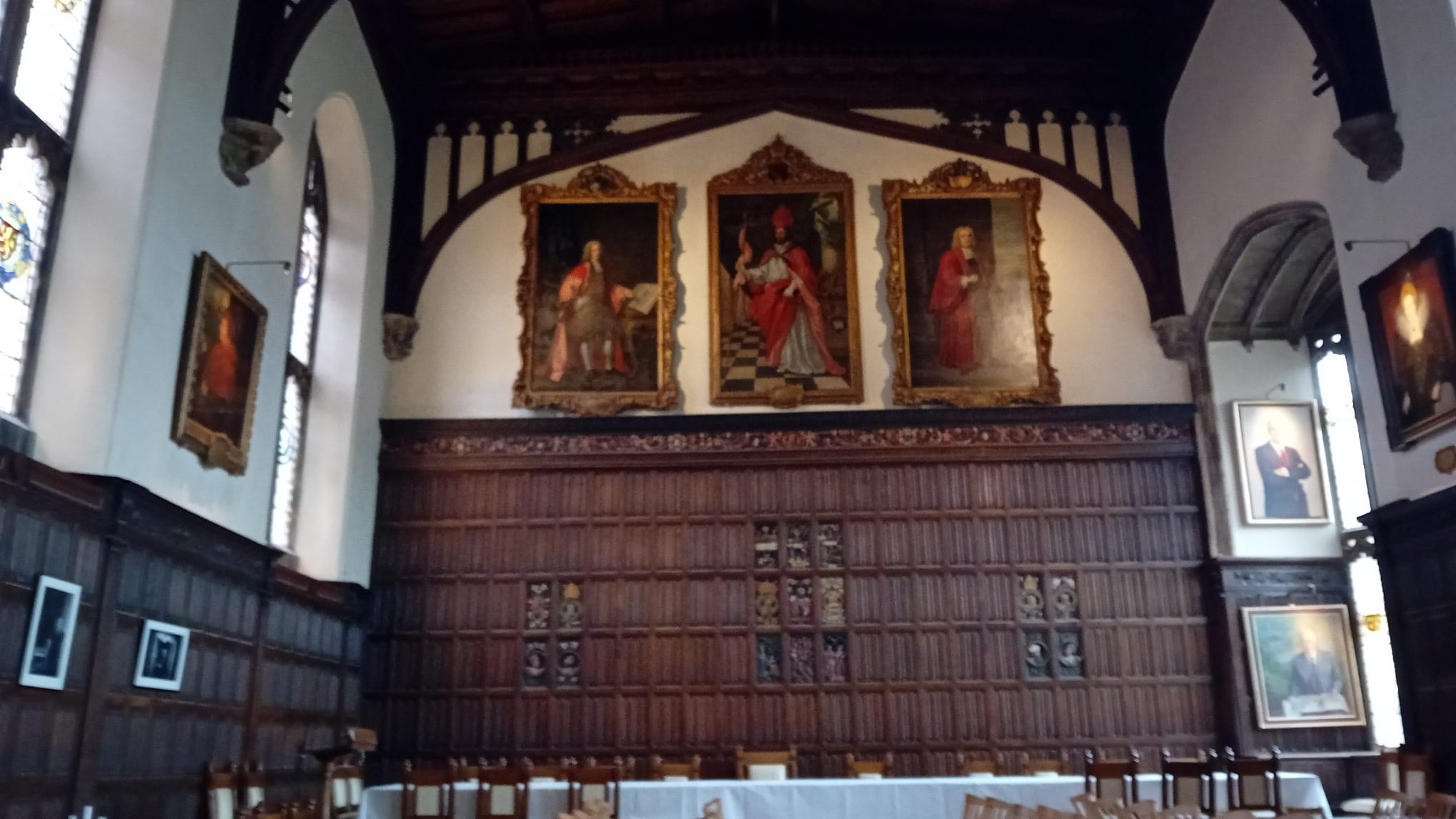
Still, it was lovely. I did have a piano in my bedroom, extremely high ceilings, the breakfast was good, and I set off with a spring in my step to the first of my five booked lectures.
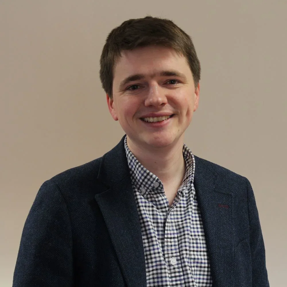
My first lecture was a refresher of the history of brain imaging, followed by information about what research using brain imaging is like now. Laurence Hunt, Professor of Cognitive Neuroscience and Associate Director of the Oxford Centre for Integrative Neuroimaging, presented this particular talk. Forget about policemen, you know you’re getting really old when even professors start looking young!
The History of Brain Imaging
Laurence started off with some history. 100 years ago, we deduced how the brain was working by looking at changes in behaviour when it was damaged (i.e. lesions). I reckon we are all probably familiar with the wonderfully named Phineas Gage, whose left frontal lobe was partially destroyed by an iron rod back in 1848. His behaviour changed in various ways – he became rude, indecisive, irritable, but many other functions remained entirely intact. He even gained employment as a stagecoach driver in Chile. We realised that different regions were responsible for different functions.
In 1924 Hans Berger began recording electrical activity by placing electrodes on the scalp – this was the EEG, and now we could detect brain waves.
In 1968 David Cohen at MIT began to measure weak magnetic fields using MEG (magnetoencephalography) in a very “sixties” type of development.
Then Wilder Penfield began experiments during surgery on epileptic patients, stimulating different regions of the cortex, and seeing what twitched, or asking what the patient experienced. He did a lot of mapping of brain areas (and produced the homunculus – that famous picture of the little man in the brain with enormous hands and lips and genitalia).
Then – Hallelujah! – in the 1970s and 1980s we began to image the living brain with MRIs and PET scans.
(MRI scans
1) use a magnet to make the nuclei of hydrogen atoms line up
2) the scanner then sends radio frequencies disturbing that alignment
3) as they return to the aligned state they release energy as radio signals.
4) The scanner converts these into an image.
So now you know.)
So what is “Functional MRI”
Everything seems to be “functional” now – we have functional nutritionists, functional medicine, functional exercise – but in the 1990s, we discovered how to use MRI scans to measure small local changes in blood flow at the area of neural activity. So when you see those images which show little patches of the brain “lighting up”, this is what you are seeing – changes in the blood flow and oxygenation of that area. This is what is known as functional MRI, often written as fMRI.
The very first data from Oxford, in the John Radcliffe, was of a right finger tapping at 2Hz (twice a second). Someone lay in an MRI scanner, wiggled their finger, and Hey Presto! there was a lighting up of the contralateral motor cortex. This was in 1998.
For the science bods amongst you, magnets seem to be measured in Teslas. (I don’t think this is connected to Elon Musk – he is more the car end of the Tesla name.) The Oxford research MRI used to have a 3 Tesla magnet. The ones they currently use have a 7 Tesla magnet i.e. much more powerful.
Functional MRI has thrown up some interesting data. For example, when they look at amputees, they discover that brain organisation can be retained long after losing afferent input from a missing limb. Kikkert et al, 2016, showed a surprising stability in the brain function even when someone had had a hand amputated.
What about now, and what is diffusion MRI?
For a long time WHERE? was the question, Laurence told us. But now in 2026 we are asking HOW?, WHY? IN WHOM? and WHAT CAN WE DO?
Through all of this research we now have a quite dizzying array of things to measure and monitor the brain with – lesions, scans, EEGs, MEGs – they look at regions of the brain, and maybe circuits. However at the cellular and molecular level we need optical imaging, microscopy, single neuron recording and transcriptomics. Some of these are only available in animals. (I did notice, during this day, across all the lectures, there was occasional reference to animals being used in experiments – mentioned but slightly glossed over. Monkeys and mice were the animals in question, with assurances that their use was minimal but incredibly useful. No real detail.)
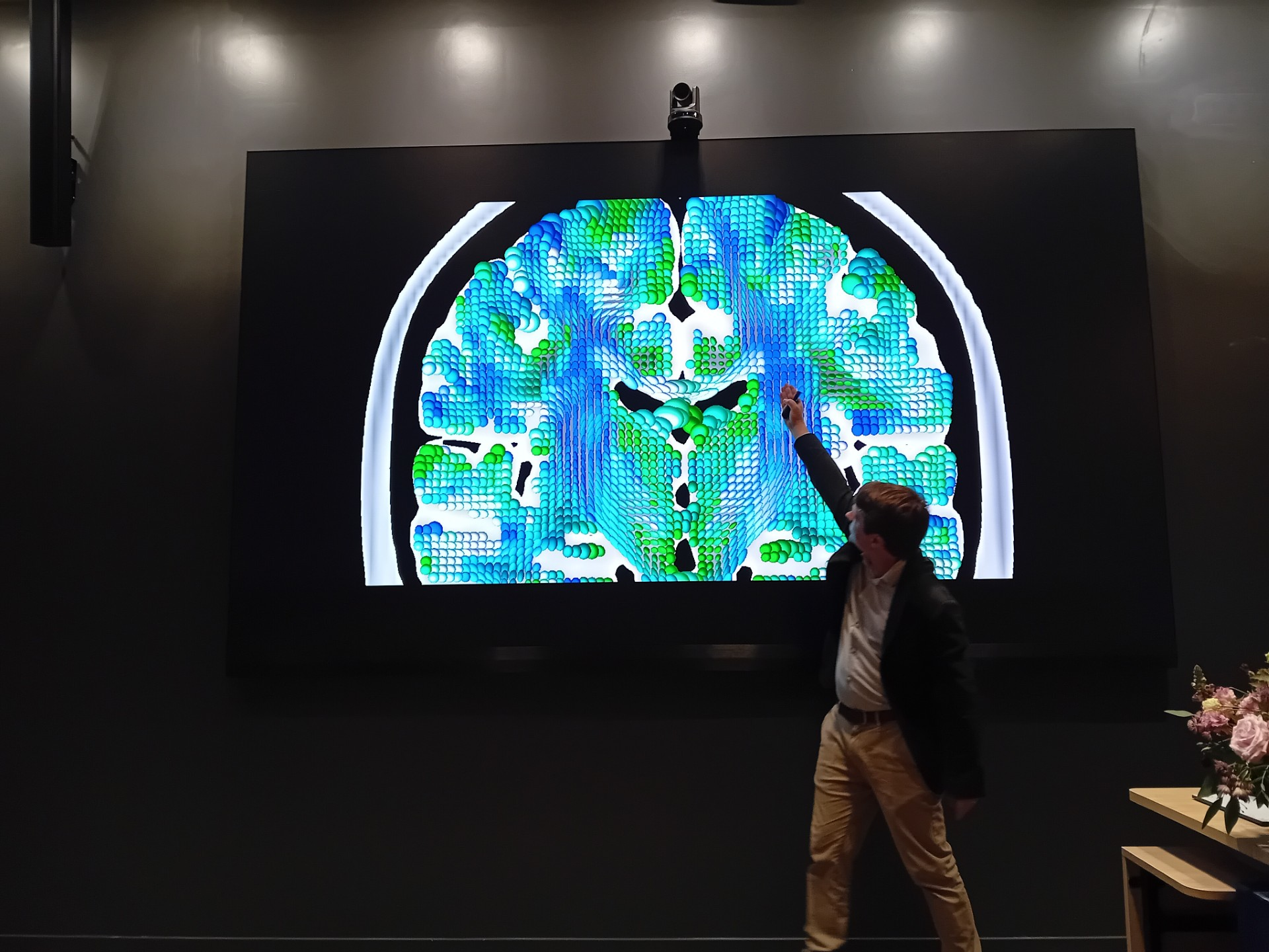
And we also have a new kid on the block – Diffusion MRI. Functional MRI shows you what the brain is doing, but diffusion MRI helps show how the brain is wired. It does this by showing the direction that water is diffusing. Because water diffuses more easily along the white tracts than across them (remember your physiology – the axons that transmit nerve impulses are the white tracts). So you can see the paths of nerve connections, and this is called “tractography”.
The pictures are different and quite psychedelic, appropriately, considering we can now see images showing the changing brain. (Psyche – delic)
What use is it?
This has therapeutic value. One can now target a specific area for treatment in the individual. For example, finding the correct area in a particular person in order to implant electrodes for deep brain stimulation in just the right place to treat the tremor of Parkinson’s.
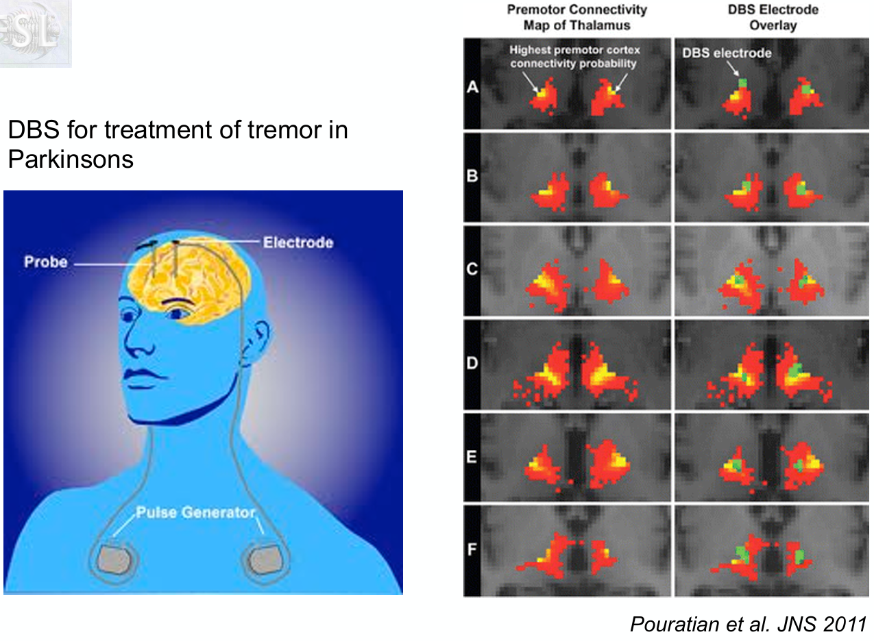
The brain varies individual to individual, he told us. There is no average brain. Currently researchers are utilizing the enormous sample sizes in the UK Biobank (see my previous blog) – 100 000 in the imaging arm – to examine the brains of 2000 people diagnosed with Alzheimer’s over 5 years. Can they pick up early markers? We wait in anticipation.
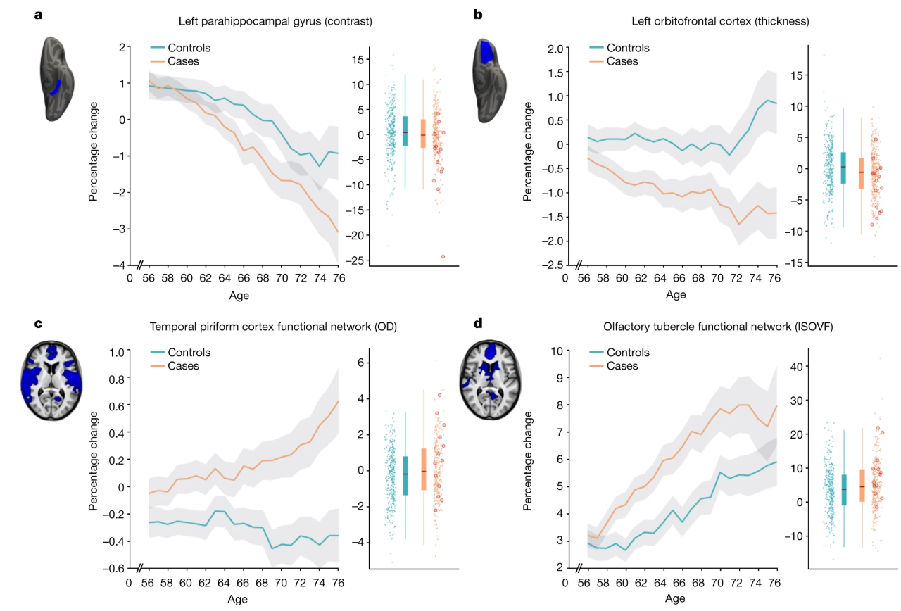
Covid brain. Many people seem to have had symptoms of this, lasting quite a while. Well, luckily for us, 401 people in the UK Biobank happened to contract covid between their 2 routine scans. And yes, we could see that they had had observable brain changes after covid. Diffusion tractography was used to study these brains and found potentially large scale structural changes, maybe including the olfactory pathway (Douad et al, 2022). They will continue to follow this up, to see if they can establish just how long covid brain lasts. I personally haven’t been able to enjoy a cup of tea quite as fully as I used to, and this might be something happening at the brain level.
Are there implications for research into osteopathy?
Obviously this might seem exciting to those osteopaths who use “cranial” techniques. Surely the Holy Grail would be to actually observe what happens during a CV4, or a frontal lift?
Functional MRI has already shown changes in connectivity following standard “osteopathic manipulative treatment” in 30 healthy young adults.(Tramontano et al, 2020, Brain Science). Maybe we will be able to scan the brain while treating and see what effect it has? I’m not even sure if you can MRI people while treating, but even before and afters are useful. Maybe the more advanced and accessible brain imaging is, we might be able to track exactly what is happening and what is changing when we have hands-on.
Where to now?
There is a move away from what the brain looks like to how it changes over time. Plasticity and degeneration are key areas of interest. Research is still somewhat hampered by being done while the subjects are in the artificial environment of the MRI scan, but some new MEG devices have flexible helmets to try to replicate real world states in their research. In addition, it is quite difficult to get data on babies, both newborn and intrauterine, for obvious wiggle and access reasons.
Questions from the audience
Can you see ADHD in a brain? Great question, prompting a lot of nervous fidgeting (in me, at least). Well, there are some changes in anticipating what sensory input you’re going to get. So we can see a predictive processing change.
Can the imaging be used for assessing drug interventions? Yes, and it is.
Final question to Laurence, a very brainy person himself also with a huge knowledge of brains, what do we do to keep our brain healthy? Well, good sleep and exercise. Again this seemed to be a repeating theme throughout the day – whichever lecture I went to, however cutting edge and advanced the research was, much of the advice seemed to come down to wholesome living.
This was a great talk – full of fascinating facts and a fab overview of brain imaging. Many thanks to Professor Hunt and his similarly brainy team for doing this valuable work, from which we will all benefit. Thanks also to checking this through for errors and giving me the go ahead/
And finally, thanks to you who are reading this. I hope you are enjoying learning as much as me!


Another entertaing and informative read. Thank you.
LikeLike